Research - (2022) Volume 13, Issue 4
Abstract
Purpose: The study investigated Interleukin-6 expression pattern across all stages of cancer. The research questions raised in the study were: Is there differential expression of Interleukin-6 across all cancer stages? W hat relationship exists between serum Interleu level and cancer stage?
Methods: The prospective case-control study comprised sixty two (62) purposively selected cancer participants across all stages and age range 18 years to 72 years as well equal number of healthy volunteers from two medical centers in Nigeria. Three milliliters (3 ml) of blood samples was collected intravenously from the participants and centrifuged after 30 minutes of collection at 3000 rpm for 10 minutes to obtain serum. The serum level of Interleukin-6 was determined spectrophotometrically by Enzyme Linked Immunosorbent Assay (ELISA). Data obtained were expressed as mean and standard error of the mean. One way Analysis of variance and t-test were employed to test for significance difference between the groups and the significant level was considered at P<0.05.
Results: Findings from the study revealed significant (P<0.05) higher mean serum Interleukin-6 levels in stage IV cancer participants as compared to other disease stages. In the same way, significant higher mean Interleukin-6 level of stage III cancer participants as compared to that of stage I cancer participants was observed. Furthermore, the study revealed a significant correlation (P<0.01) between serum Interleukin-6 concentration and cancer stage.
Conclusion: Serum Interleukin-6 had differential expression in cancer patients at advanced clinical stage as compared to that of early disease stage.
Keywords
Interleukin, Spectrophotometry, Enzyme Linked Immunosorbent Assay (ELISA), Cancer, Centrifuge, Prospective
Introduction
Cancer, a public health problem afflicting all categories of person is among the global issues, with an estimated 10 million incidences and 6 million annual mortality rates (Abdulkareem F, 2009). It is a group of diseases that are characterized by uncontrolled cellular growth, local tissue invasion and distant metastasis caused by aberrations in cellular regulatory mechanisms (Aggarwal BB and Gehlot P, 2009).
Staging of cancer is an important process in cancer evaluation and clinical treatment of solid tumors in which the extent of tumor growth and its spread in the body system are revealed. TNM (Tumor, Nodes, Metastasis) system is one of the most widely adopted methods of cancer staging which employs histopathological evaluations to assign stages between I and IV on the basis of primary tumor size (T), degree of lymph node involvement (N) and the extent of metastasis (M) (Angahar LT, 2017; Akinde OR, et al., 2014).
Recently, there has been an increasing interest in cancer-related inflammation as a major determinant of disease progression and survival in the field of oncology research (Belluco C, et al., 2000; Chung YC and Chang YF, 2003). Numerous studies provide evidence that chronic inflammation increases cancer risk, promote tumor progression and support metastasis (de Vita FE, et al., 1998; Kodati D, et al., 2019).
Interleukin-6 has been subject to increased attention due to its possible usefulness as a biomarker of inflammation (Ghandadi M and Sahebkar A, 2016). The strong association between inflammation mediated Interleukin-6 and cancer development and progression has been well documented (Mittal P, et al., 2016; Weinberg RA and Hanahan D, 2000).
Owing to the fact that inflammatory signaling plays key roles in cancer development, progression and therapy response (Ghandadi M and Sahebkar A, 2016), determination and comparison of serum Interleukin-6 expression pattern across cancer patients of different disease stage might reveal a better and more reliable predictive prognostic marker as well as identifying target oncogenic mediator that will be of therapeutic importance for the prevention of cancer progression and improved survival outcomes of cancer patients. Therefore, the study investigated Interleukin-6 expression pattern in cancer patients of different disease stage from selected Nigeria tertiary health care.
Materials and Methods
Experimental design and participants
The prospective case-control study comprised sixty two (62) purposively selected cancer participants across all stages and age range 18 years to 72 years as well equal number of healthy volun teers from two medical centers in Nigeria, namely: University College Hospital (UCH), and Federal Medical Centre (FMC), Idi- aba, Abeokuta.
Participants were included in the study on the basis of absence of neurological disorders and infection, no history of opoid use and they were not on immunosuppressive drugs and anti-inflammatory drugs 24 hrs prior to the study. However, Patients with incidence of pre-existing acute or chronic inflammatory diseases or infectious diseases as well as apparent multiple organ dysfunction were excluded from the study.
Ethical approval
The approval of the study was obtained from ethics review committee of the selected medical centers with the following ethics committee assigned numbers; UI/EC/19/0161 and FMCA/470/HREC/01/2019/010 respectively. The procedure followed for the conduction of the study was in accordance with the ethical standards of the selected medical institution and with the revised Helsinki Declaration of 1975.
Also, every participant gave their consent prior to the conduction of the study by appending their signatures on the informed consent form.
Participant study
Socio-demographics of the consented participants were collected from the patients using a well-structured questionnaire.
Clinical features of the participants
Participants’ clinical information as regard to their cancer stage was retrieved from the hospital. The cancer staging of the participants was based on “TNM” system.
Sample collection
Three milliliters (3 ml) of blood samples was collected intravenously from the cancer subjects into a set of well labeled plain sample bottles. The collected blood samples were left standing for 30 minutes after which it was centrifuged at 3000 rpm for 10 minutes for serum separation. The collected serum was kept in a bio freezer set at -20°C until sample analysis.
Enzyme Linked Immunosorbent Assay (ELISA)
The expression levels of Interleukin-6 cytokines in the separated sera samples were determined spectrophotometrically by Enzyme Linked Immunosorbent Assay using Interleukin-6 specified ELISA kit as described by Mittal P, et al. (Hanahan D and Weinberg RA, 2011).
Statistical analysis
All experiments were set up in triplicates. Data collected were processed using SPSS statistical software version 22.0 and the processed data were expressed as mean ± standard deviation. Analysis of variance and paired t-test were employed to test for significance difference among the variables with the level of significance considered at P<0.05.
Results
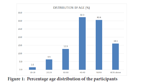
Figure 1 shows the percentage age distribution of the participants selected for the study. It was evident from Figure 1 that majority of the cancer patients that participated in the study were of the age groups 40-49 years and 50-59 years with percentage distribution of 32.3% and 30.6% respectively. While minority of the study group comprised of cancer patients of the age group 10- 19 years with percentage distribution of 1.6%.

Figure 1:Percentage age distribution of the participants
The percentage of different types of cancer patients included in the study was presented in Table 1. From Table 1, majority of the participants were breast cancer and cervical cancer patients as it was reflected in their percentage distribution of 48.4 and 17.7% respectively. However, patients with neck cancer, brain cancer, Non Hodgkin lymphoma, prostate, Rectal and skin cancer constituted the minority of the participants with percentage distribution of 1.6%.
| Disease subtype | Frequency | Percentage(%) |
|---|---|---|
| Ameloblastic carcinoma | 3 | 4.8 |
| Brain cancer | 1 | 1.6 |
| Breast cancer | 30 | 48.4 |
| Cervical cancer | 11 | 17.7 |
| Eye cancer | 1 | 1.6 |
| Larynx cancer | 2 | 3.2 |
| Maxillary adenoid carcinoma | 2 | 3.2 |
| Nasopharyngeal cancer | 2 | 3.2 |
| Neck cancer | 1 | 1.6 |
| Non hodgkin lymphoma | 1 | 1.6 |
| Oropharyngeal cancer | 3 | 4.8 |
| Prostate cancer | 1 | 1.6 |
| Rectal cancer | 1 | 1.6 |
| Skin cancer | 1 | 1.6 |
| Tongue cancer | 2 | 3.2 |
| Total | 62 | 100 |
Table 1: Percentage distribution of the participants by cancer types
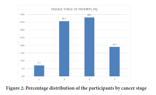
Figure 2 revealed the percentage of the cancer patients in different stages of the disease. The figure showed that higher population of the participants involved in the study were in stage 2 and stage 3 of the disease as it was reflected in their percentage distribution of 35.7% and 38.1% respectively. However, cancer patients at stage 1 of the disease had the lowest percentage (7.1%) of all the recruited participants.

Figure 2:Percentage distribution of the participants by cancer stage
The mean serum Interleukin-6 levels of cancer participants and healthy volunteers were revealed in the Table 2. As revealed from the table, the mean serum Interleukin-6 levels of the cancer patients was significantly (P<0.05) higher than that of the control.
| Participants | Mean serum Interleukin-6 level | P value |
|---|---|---|
| Cancer patients | 84.15 ± 16. 55* | 0.016 |
| Control | 61.56 ± 7.144 |
Note: values with *indicates significance (P<0.05) difference in Interleukin-6 concentration
Table 2: Mean serum Interleukin-6 level of the participants
The serum mean Interleukin-6 concentration of the participants with different stages of cancer is presented in Table 3 above. From the table, the mean Interleukin-6 concentration increases with advancing stage of disease. However, stage IV participants (with metastatic disease) have significant (P<0.05) higher concentration of Interleukin-6 as compared to other stages. In the same way, significant increase in the mean Interleukin-6 concentration of participants with stage III of the disease as compared to that of stage I cancer participants was evident from Table 3.
| Cancer stage group | Mean Interleukin-6 concentration |
|---|---|
| I | 15.76 ± 10.04a |
| II | 20.64 ± 5.13ab |
| III | 45.31± 5.08b |
| IV | 174.20 ± 46.14c |
Note: Values with different alphabets superscript indicate significance difference (P<0.05) in Interleukin-6 concentration
Table 3: Mean values of Serum Interleukin-6 concentration of the participants of different disease stage
Interleukin-6 level in the serum of the participants with different cancer types was revealed as in Table 4. From the table, there was a significant decrease (P<0.05) in the serum Interleukin-6 level of breast cancer participants as compared to that of cervical, prostate, head and Neck and others cancer types.
| Cancer type | Serum Interleukin-6 level |
|---|---|
| Breast Cancer | 24.311 ± 6.33* |
| Cervical Cancer | 160.53 ± 47.70 |
| Prostate Cancer | 49.53± 6.432 |
| Head and Neck Cancer | 73.60 ± 26.02 |
| Others | 89.53 ± 39.13 |
Note: Value with *indicates significance difference (P<0.05) in Interleukin-6 level
Table 4: Mean values of Serum Interleukin-6 levels of the participants with different cancer types
From Table 5 above, there was a significant correlation (P<0.01) between Interleukin-6 concentration and cancer age.
| Mean serum Interleukin 6 level | Pearson correlation coefficient | P value |
|---|---|---|
| Interleukin-6 levels | 1 | 0.002* |
| Cancer stage | 0.466* |
Note: *Correlation is significant at the 0.01 level (2-tailed)
Table 5: Correlation relationship between Interleukin-6 concentrations and cancer stage
Discussion
Of recent, there has been an increasing interest in cancer-related inflammation as a major determinant of disease progression and survival in the field of oncology research. Interleukin-6 (IL6) has been implicated as a member of the major inflammatory Interleukins family that has played a significant role in various immune, physiological and pathophysiological processes such as cancer progression (Hunter CA and Jones SA, 2015; Jedy-Agba E, et al., 2012; Jeffrey SS, et al., 1999; Kozłowski L, et al., 2003). Determination of the expression pattern of Interleukin-6, an important inflammatory marker, might reveal a reliable predictive prognostic marker as well as identifying target oncogenic mediator that will be of therapeutic importance for the prevention of cancer progression and improves survival outcomes of cancer patients. Therefore, the present study investigated Interleukin-6 expression pattern in cancer patients with different disease stages from selected Nigeria tertiary health care.
Findings from the participants’ socio-demographics analysis of this present study revealed Breast cancer and cervical cancer as most common cancer of all the cancer types among the studied participants. This findings corroborates the findings of Jedy-Agba E, et al. in which Breast and cervical cancer were reported as the most common cancers in women from the study conducted in two regions in Nigeria (Table 1) (Kumari N,et al., 2016). Also, this finding is in line with the epidemiological and incidence report of common cancers in Nigeria by Abdul kareem F in which Breast and Cervical cancer were revealed as first two most common cancers in Nigeria (Lederle W, et al., 2011).
Considering the age distribution of the included participants, age group 40-49 years was revealed from the findings of the study as the most vulnerable age group to cancer disease (Figure 1). The findings obtained is in line with the work (Mantovani A, et al., 2008) on cancer mortality pattern in Lagos University Teaching Hospital, in which age group 41-50 were reported as the most cancer prevalent age groups with percentage distribution of 21.0% respectively (Mantovani A, et al., 2008). In light of the above, advanced age is an important risk factor of cancer development. Descriptive survey on the cancer stage of the participants in this present study revealed that highest proportions of the participants were at advanced stage (III) of the disease while only small proportion of the studied population were at early stage of cancer. Possible reasons for this observation might be due to late diagnosis and late presentation in the hospitals for cancer treatment.
For the serum Interleukin 6 levels, findings from this present study revealed that Interleukin-6 levels in the serum of the participants diagnosed with advanced cancer stage (III and IV) was significantly elevated as compared to that of early stage of the disease. This agrees with earlier reports from a number of studies conducted previously which showed that Patients, who have Cancer have increased levels of IL-6 (Masjedi A, et al., 2018; Nakagoe T, et al., 2003; Vainer N, et al., 2018; Okamoto M, et al., 1997).
Reports from previous studies suggest that an increased production of IL-6 is associated with poor prognosis and survival outcomes in many types of cancers including Breast Cancer (Roche K, et al., 2017; Shibayama O, et al., 2014). Furthermore, our study showed that IL-6 levels were highest in Cancer patients at stage 4, when compared to the other stages in all the Cancer types. Similarly, Chung YC and Chang YF reported that an existing association occurred between colorectal tumor size and IL-6 serum levels which peaked at Stage IV in the Patients having colorectal cancer (Taher MY, et al., 2018).
Conclusion
Conclusively, serum Interleukin-6 differential expression in cancer patients at advanced clinical stage as compared to that of early disease stage, alongside with the significant association relationship between its expression level and the different clinical stages of cancer as evident from the concluded study. Hence, serum Interleukin-6 is highlighted as a promising biomarker of tumor progression and prognosis as well as an important therapeutic target to prevent tumor progression and metastasis for improved survival outcomes of cancer patients.
Acknowledgement
This research was supported by Tertiary Education Trust (TETFUND) Grant with document code TETFUND/DESS/FCE/OSIELE/2017/RP/ VOL I, and the authors hereby express gratitude to TETFUND for this.
References
- Abdulkareem F. Epidemiology and incidence of common cancers in Nigeria. Cancer Reg and Epid. 2009.
- Aggarwal BB, Gehlot P. Inflammation and cancer: How friendly is the relationship for cancer patients? Curr Opin Pharmacol. 2009; 9(4): 351-369.
[Crossref] [Google Scholar] [Pubmed]
- Akinde OR, Phillips AA, Oguntunde OA, Afolayan OM. Cancer mortality pattern in Lagos University teaching hospital, Lagos, Nigeria. J Cancer Epidemiol. 2014; 2015.
[Crossref] [Google Scholar] [Pubmed]
- Angahar LT. An overview of breast cancer epidemiology, risk factors, pathophysiology, and cancer risks reduction. MOJ Biol Med. 2017; 1(4): 19.
- Belluco C, Nitti D, Frantz M, Toppan P, Basso D, Plebani M, et al. Interleukin-6 blood level is associated with circulating carcinoembryonic antigen and prognosis in patients with colorectal cancer. Ann Surg Oncol. 2000; 7(2): 133-138.
[Crossref] [Google Scholar] [Pubmed]
- Chung YC, Chang YF. Serum interleukin‐6 levels reflect the disease status of colorectal cancer. J Surg Oncol. 2003; 83(4): 222-226.
[Crossref] [Google Scholar] [Pubmed]
- de Vita FE, Orditura MI, Auriemma AN, Infusino ST, Roscigno AN, Catalano GI. Serum levels of interleukin-6 as a prognostic factor in advanced non-small cell lung cancer. Oncol Rep. 1998; 5(3): 649-701.
[Crossref] [Google Scholar] [Pubmed]
- Kodati D, Tirumala S, Kantheti RK, Thumma JP. A retrospective study on the adverse effects of chemotherapy in breast cancer-a multicenter study. Indian J Pharm Pract. 2019; 12(2): 111.
- Ghandadi M, Sahebkar A. Interleukin-6: A critical cytokine in cancer multidrug resistance. Curr Pharm Des. 2016; 22(5): 518-526.
[Crossref] [Google Scholar] [Pubmed]
- Mittal P, Gupta N, Goswami B. Serum IL-6 level as a predictor of response to neo-Adjuvant chemotherapy in patients of breast carcinoma. Hellenic J Cardiol. 2016; 88(5): 306-310.
- Weinberg RA, Hanahan D. The hallmarks of cancer. Cell. 2000; 100(1): 57-70.
[Crossref] [Google Scholar] [Pubmed]
- Hanahan D, Weinberg RA. Hallmarks of cancer: The next generation. Cell. 2011; 144(5): 646-674.
[Crossref] [Google Scholar] [Pubmed]
- Hunter CA, Jones SA. IL-6 as a keystone cytokine in health and disease. Nat Immunol. 2015; 16(5): 448-457.
[Crossref] [Google Scholar] [Pubmed]
- Jedy-Agba E, Curado MP, Ogunbiyi O, Oga E, Fabowale T, Igbinoba F, et al. Cancer incidence in Nigeria: A report from population-based cancer registries. Cancer Epidemiol. 2012; 36(5): 271-278.
[Crossref] [Google Scholar] [Pubmed]
- Jeffrey SS, Birdwell RL, Ikeda DM, Daniel BL, Nowels KW, Dirbas FM, et al. Radiofrequency ablation of breast cancer: First report of an emerging technology. Arch Surg. 1999; 134(10): 1064-1068.
[Crossref] [Google Scholar] [Pubmed]
- Kozłowski L, Zakrzewska I, Tokajuk P, Wojtukiewicz MZ. Concentration of interleukin-6 (IL-6), interleukin-8 (IL-8) and interleukin-10 (IL-10) in blood serum of breast cancer patients. Rocz Akad Med Bialymst. 2003; 48: 82-84.
[Google Scholar] [Pubmed]
- Kumari N, Dwarakanath BS, Das A, Bhatt AN. Role of interleukin-6 in cancer progression and therapeutic resistance. Tumor Biol. 2016; 37(9): 11553-11572.
[Crossref] [Google Scholar] [Pubmed]
- Lederle W, Depner S, Schnur S, Obermueller E, Catone N, Just A, et al. IL‐6 promotes malignant growth of skin SCCs by regulating a network of autocrine and paracrine cytokines. Int J Cancer. 2011; 128(12): 2803-2814.
[Crossref] [Google Scholar] [Pubmed]
- Mantovani A, Allavena P, Sica A, Balkwill F. Cancer-related inflammation. Nature. 2008; 454(7203): 436-444.
[Crossref] [Google Scholar] [Pubmed]
- Masjedi A, Hashemi V, Hojjat-Farsangi M, Ghalamfarsa G, Azizi G, Yousefi M, et al. The significant role of interleukin-6 and its signaling pathway in the immunopathogenesis and treatment of breast cancer. Biomed Pharmacother. 2018; 108: 1415-1424.
[Crossref] [Google Scholar] [Pubmed]
- Nakagoe T, Tsuji T, Sawai T, Tanaka K, Hidaka S, Shibasaki SI, et al. Increased serum levels of interleukin-6 in malnourished patients with colorectal cancer. Cancer Lett. 2003; 202(1): 109-115.
[Crossref] [Google Scholar] [Pubmed]
- Vainer N, Dehlendorff C, Johansen JS. Systematic literature review of IL-6 as a biomarker or treatment target in patients with gastric, bile duct, pancreatic and colorectal cancer. Oncotarget. 2018; 9(51): 29820.
[Crossref] [Google Scholar] [Pubmed]
- Okamoto M, Lee C, Oyasu R. Interleukin-6 as a paracrine and autocrine growth factor in human prostatic carcinoma cells in vitro. Cancer Res. 1997; 57(1): 141-146. [Crossref]
[Google Scholar] [Pubmed]
- Roche K, Feltus FA, Park JP, Coissieux MM, Chang C, Chan VB, et al. Cancer cell redirection biomarker discovery using a mutual information approach. PloS One. 2017; 12(6): 179265.
[Crossref] [Google Scholar] [Pubmed]
- Shibayama O, Yoshiuchi K, Inagaki M, Matsuoka Y, Yoshikawa E, Sugawara Y, et al. Association between adjuvant regional radiotherapy and cognitive function in breast cancer patients treated with conservation therapy. Cancer Med. 2014; 3(3): 702-709.
[Crossref] [Google Scholar] [Pubmed]
- Taher MY, Davies DM, Maher J. The role of the interleukin (IL)-6/IL-6 receptor axis in cancer. Biochem Soc Trans. 2018; 46(6): 1449-1462.
[Crossref] [Google Scholar] [Pubmed]
Author Info
Bello AO1*, Obadipe JA1,2, Adewusi OA2 and Ayanshina AO22Department of Biochemistry, College of Medicine, University of Lagos, Lagos State, Nigeria
Citation: Bello AO: Interleukin-6 Differential Expression in Cancer Patients of Different Clinical Stages: A Possible Biomarker of Cancer Progression
Received: 08-Mar-2022 Accepted: 29-Mar-2022 Published: 05-Apr-2022, DOI: 10.31858/0975-8453.13.4.265-268
Copyright: This is an open access article distributed under the terms of the Creative Commons Attribution License, which permits unrestricted use, distribution, and reproduction in any medium, provided the original work is properly cited.
ARTICLE TOOLS
- Dental Development between Assisted Reproductive Therapy (Art) and Natural Conceived Children: A Comparative Pilot Study Norzaiti Mohd Kenali, Naimah Hasanah Mohd Fathil, Norbasyirah Bohari, Ahmad Faisal Ismail, Roszaman Ramli SRP. 2020; 11(1): 01-06 » doi: 10.5530/srp.2020.1.01
- Psychometric properties of the World Health Organization Quality of life instrument, short form: Validity in the Vietnamese healthcare context Trung Quang Vo*, Bao Tran Thuy Tran, Ngan Thuy Nguyen, Tram ThiHuyen Nguyen, Thuy Phan Chung Tran SRP. 2020; 11(1): 14-22 » doi: 10.5530/srp.2019.1.3
- A Review of Pharmacoeconomics: the key to “Healthcare for All” Hasamnis AA, Patil SS, Shaik Imam, Narendiran K SRP. 2019; 10(1): s40-s42 » doi: 10.5530/srp.2019.1s.21
- Deuterium Depleted Water as an Adjuvant in Treatment of Cancer Anton Syroeshkin, Olga Levitskaya, Elena Uspenskaya, Tatiana Pleteneva, Daria Romaykina, Daria Ermakova SRP. 2019; 10(1): 112-117 » doi: 10.5530/srp.2019.1.19
- Dental Development between Assisted Reproductive Therapy (Art) and Natural Conceived Children: A Comparative Pilot Study Norzaiti Mohd Kenali, Naimah Hasanah Mohd Fathil, Norbasyirah Bohari, Ahmad Faisal Ismail, Roszaman Ramli SRP. 2020; 11(1): 01-06 » doi: 10.5530/srp.2020.1.01
- Manilkara zapota (L.) Royen Fruit Peel: A Phytochemical and Pharmacological Review Karle Pravin P, Dhawale Shashikant C SRP. 2019; 10(1): 11-14 » doi: 0.5530/srp.2019.1.2
- Pharmacognostic and Phytopharmacological Overview on Bombax ceiba Pankaj Haribhau Chaudhary, Mukund Ganeshrao Tawar SRP. 2019; 10(1): 20-25 » doi: 10.5530/srp.2019.1.4
- A Review of Pharmacoeconomics: the key to “Healthcare for All” Hasamnis AA, Patil SS, Shaik Imam, Narendiran K SRP. 2019; 10(1): s40-s42 » doi: 10.5530/srp.2019.1s.21
- A Prospective Review on Phyto-Pharmacological Aspects of Andrographis paniculata Govindraj Akilandeswari, Arumugam Vijaya Anand, Palanisamy Sampathkumar, Puthamohan Vinayaga Moorthi, Basavaraju Preethi SRP. 2019; 10(1): 15-19 » doi: 10.5530/srp.2019.1.3


