Research Article - (2023) Volume 14, Issue 3
Abstract
Objective: Primary brain tumors are classified as glial or non-glial and benign or malignant. Meningiomas are common benign intracranial tumors. Although the name meningioma refers to a tumor of the lining of the brain called the ‘Meninx’, it has actually been shown to originate from the spider web-shaped ‘arachnoid’ membrane (arachnoid cover cells). microRNAs are 18- 22 nucleotide long, endogenous, non-protein-coding RNA molecules that negatively regulate gene expression at the post-transcriptional level. In this study, we applied a genome-wide array screen comparing the expression of miR-145, miR-34a-3p, miR-200a, miR- 335, miR-106a-5p, miR-219-5p, miR-375, miR-409-3p miR-197 and miR-224 in meningiomas.
Materials and methods: A total of 40 meningioma patients (13 men, 27 women) and healthy control individuals (12 men, 18 women) aged between 30 and 65 were inclusives in the study. The research was conducted at Gazi University Hospital.
Results: In our study, miR-197 identified as the most highly expressed miRNA in meningiomas compared to other miRNAs. miR-197, miR-34a, miR-375, miR- 219a and miR-224 stand out as potential biomarkers in human serum samples of meningiomas patients. Moreover, as per WHO classification miR-197, miR- 34a, miR-375 might be used as potential biomarkes for grade I meningioma while miR-375 for grade II meningioma.
Conclusion: The role of miRNAs in meningiomas is gaining importance each day. Therefore, our study examining the role of miRNAs in meningiomas will shed more light and pave the way for future therapeutic strategy.
Keywords
Biomarker, Meningioma, miRNA, miR-197, miR-34a-3p
Introduction
Meningiomas can diffuse Central Nervous System (CNS) tumors that account for approximately one-third of all intracranial tumors (Galani V, et al., 2017; Dolecek TA, et al., 2012). According to the World Health Organization (WHO) classification, the vast majority of meningiomas are grade 1 (typical/benign), less than 10% grade II (atypical/moderate) and III (anaplastic/malignant) tumors (Mei Y, et al., 2017). Their incidence increases with age with a Female:Male ratio of 2:1. Furthermore, findings from autopsy and imaging studies show that the prevalence of subclinical meningiomas constitutes roughly 3% of the population (SöylEmEzoğlu F, 2011). It has been implicated in the pathogenesis of many tumors, such as inactivation of tumor suppressor genes or overexpression of oncogenes and specific gene dysfunctions (Amplifikasyonu G, 2009). The most common abnormality identified in meningiomas is deletions in chromosome 22. Loss of chromosomes 1p, 3p, 6q, 10q and 14q were also observed, especially in atypical or malignant meningiomas. It has already been suggested that these deletions can be used as a marker to predict tumor progression (Arslantas A, et al., 2022). microRNAs are 18-22 nucleotide long, endogenous, non-protein-coding RNA molecules that negatively regulate gene expression at the post-transcriptional level (McManus MT, et al., 2002). In recent years, there has been an increasing research on miRNAs as potential biomarkers for various pathological conditions, including tumors (Tie J, et al., 2010). miRNAs can function as oncogenes or tumor suppressors under certain conditions. There are evidences that they play roles in many cellular processes that contribute to tumor formation and development, from proliferation to invasion, from metastasis to angiogenesis (Tie J, et al., 2010; Vishnoi A and Rani S, 2017). Many studies analyzing the profile and function of miRNA have yielded important insights into the pathogenesis of different types of tumors, including Glioblastoma Multiforme (GBM), breast cancer, pancreatic cancer, lung cancer, prostate cancer, and colon cancer (Petrescu GE, et al., 2019; Mulrane L, et al., 2013; Uddin A and Chakraborty S, 2018; Ahmed FE, 2014; Yonemori K, et al., 2017). The recent findings indicating that miRNAs are expressed in human blood holds great promise that circulating miRNAs can serve as novel molecular biomarkers for cancer (Yonemori K, et al., 2017; Zhang K, et al., 2017). In present study, the association between meningioma and gene expression of miR-145, miR-34a-3p, miR-200a, miR-335, miR-106a-5p, miR219-5p, miR-375, miR-409-3p miR-197 and miR-224 were examined in tumor samples obtained from patients with meningioma and healthy individual samples as controls.
Materials and Methods
Study population
A total of 40 patients who applied to the Department of Neurosurgery and diagnosed with meningioma from brain tumors and 30 healthy volunteers were included in the study. Healthy group were selected from those who applied to Gazi hospital’s check-up center and whose blood sample came to the central laboratory, and those who met our admission and exclusion criteria. Some of the serums obtained from the routine blood of these people were reserved for the study. Likewise, some of the serums obtained from the blood samples taken from meningioma patients at the time of diagnosis were separated. All separated serums were stored at -80 degrees until analysis.
For the qRT-PCR assay, a fixed constant of synthetic cel-miR-39 (Genepharm, China) was added to 100 μl serum sample as the internal control. Total RNA isolation from samples was performed using the QIAGEN miRNeasy Serum/Plasma Advanced (Cat.no:217204) kit. Overall, total RNA was obtained from 200 µl serum in a yield of approximately 75-100 µng. The cDNA synthesis was made from the RNA samples using the kit (Cat.No:339340, miRCURY LNA RT Kit, Qiagen). Quantitative Reverse Transcription-Polymerase Chain Reaction (qRT-PCR) assay was performed by BioMark HD system (Fluidigm, South San Francisco, CA, USA) according to the manufacturer’s instructions. All reactions, was performed in triplicate, including controls without template RNA. The expression levels of selected miRNAs were normalized to cel-miR-39 (Zhang K, et al., 2017).
Statistical analysis
For the statistical evaluation, the conformity of the variables to the normal distribution was examined with the Shapiro-Wilk test. Normally distributed t-test was used to compare the groups. The data which were not normally distributed, Mann-Whitney U test for comparison analyzes between two independent groups, Kruskal Wallis test for comparison analyzes between three groups, post hoc test: Mann-Whitney U test with Bonferroni correction descriptive statistics are expressed as Mean ± SE and Mean ± SD. One-Way Anova:Post-hoc:Tukey HSD Test, tamhane t2 test, Dunnett test, Kruskal Wallis H test, Post hoc:Dunn Sidak tests were used to compare the expression levels of miRNAs. Statistical significance was accepted as p<0.05. IBM SPSS package program version 22 was used in the evaluation of the data.
Results
A total of 40 patients who applied to the Department of Neurosurgery and diagnosed with meningioma from brain tumors and 30 healthy volunteers were included in the study. Having 40 meningioma patients with a mean age of 48.77 ± 13.09 and 30 healthy controls with a mean age of 44.34 ± 15.04 years. A total of 70 participants were included in this study (Table 1).
| Groups | Age | Sex | |
|---|---|---|---|
| Male | Female | ||
| (Mean ± SD) | (Mean ± SD) | (Mean ± SD) | |
| Meningiom | 48.77 ± 13.9 | 46.92 ± 10.9 (n:13) |
49.81 ± 11.4 (n:27) |
| Healthy | 44.34 ± 15.4 | 47.72 ± 12.7 (n:12) |
40.16 ± 16.7 (n:18) |
| p-value | p>0.05 | p>0.05 | p>0.05 |
Table 1: Age and gender data of groups
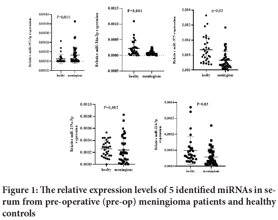
A multiphase study was designed to identify markedly altered miRNAs in serum that could distinguish meningioma from healthy controls and serve as potential biomarkers for meningioma. The study was conducted to compare the expression of human miRNAs in pooled serum samples from 40 pre-operative meningioma participants with those from 30 healthy controls for initial screening. The serum fold change of the preoperative group compared with the healthy control group is shown in Figure 1.

Figure 1: The relative expression levels of 5 identified miRNAs in serum from pre-operative (pre-op) meningioma patients and healthy controls
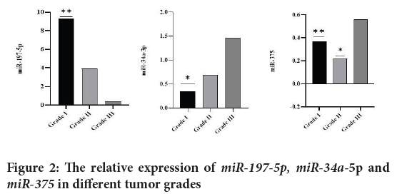
The expression levels of the 10 miRNAs in the 40 pre-operative serum samples were stratified using 3 types of clinicopathological parameters (Age, sex and WHO tumor grade). When classified according to tumor grade, miR-197-5p, miR-34a-3p and miR-375 showed significant differences associated with pathological grades (Figure 2).

Figure 2: The relative expression of miR-197-5p, miR-34a-5p and miR-375 in different tumor grades
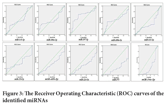
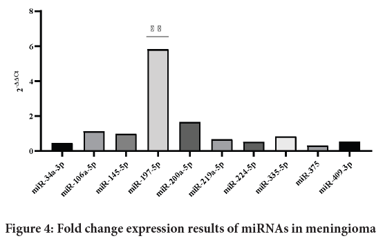
Receiver Operating Characteristic (ROC) curve analyzes were performed to evaluate the diagnostic accuracy of miRNAs in the serum between meningioma patients and normal subjects. The Area Under the Curve (AUCs) and 95% Confidence Intervals (CI) for miR-145,miR-34a-3p,miR-200a,miR-335, miR-106a-5p, miR-219-5p, miR-375, miR-409-3p miR-197 and miR-224is shown in Figures 3 and 4.
Figure 3: The Receiver Operating Characteristic (ROC) curves of the identified miRNAs
Figure 4: Fold change expression results of miRNAs in meningioma
Discussion
microRNAs play important roles in post-transcriptional gene regulation. It is known to regulate many pathways involved in cell proliferation, morphogenesis and differentiation (Vishnoi A and Rani S, 2017). Saydam O, et al., examined the relationship between different types of miRNAs and meningiomas and found that it altered miRNA levels in different types of meningiomas, with increased expression in some and decreased expression in others. They found an increase in the expression levels of miR-335, miR-98 and miR-181a, and a decrease in the expression levels of miRNA- 200a, miRNA-373 and miRNA-575 in different meningioma cases. Moreover, miRNA-200a inhibited meningioma growth during in vitro studies (Saydam O, et al., 2009). Various miRNAs (including miR-106a-5p, miR- 219-5p, miR-375, miR-197, miR-224 and miR-409-3p) are known to be dysregulated in meningioma patients (Zhi F, et al., 2016).
In previous studies, downregulation of miR-34a-3p in high-grade meningiomas has been reported (Werner TV, et al., 2017; El-Gewely MR, et al., 2016). miR-34a-3p targets the Bcl-2 gene in HBL-52 cells. It has been suggested that high expression of Bcl-2 may contribute to increased cell proliferation and decreased apoptosis of tumor cells (Ludwig N, et al., 2015). Overexpression of miR-34a-3p in meningioma inhibits cell proliferation and induces apoptosis. Therefore, it is thought that miR-34a-3p can be used as an anticancer drug in meningiomas (Misso G, et al., 2014). In our study, similar results were obtained which complements previous studies on the miR-34a-3p expression in meningiomas. We found that miR-34a-3p expression was lower in the meningioma patient group compared to the healthy control group (p<0.001).
miR-145 expression was seen in atypical and anaplastic tumours as crosschecked to benign meningiomas (Yao Z, et al., 2017). Moreover, meningioma cells with high miR-145 levels had impaired migratory and invasive potential in vitro and in vivo. Polymerase Chain Reaction (PCR) array studies of miR-145 overexpressing cells suggested that Collagen type V alpha (COL5A1) expression was down regulated as a consequence of miR- 145 over expression. Accordingly, COL5A1 expression was significantly upregulated in atypical and anaplastic meningiomas (Wilisch-Neumann A, et al., 2014).
miR-197 has been extensively studied in the carcinogenesis progression of cancers through a variety of mechanisms, including apoptosis, proliferation, angiogenesis, metastasis, drug resistance and tumor suppressor, and has also been implicated in the prognosis of cancers (Wang DD, et al., 2016; Dai W, et al., 2014; Ghafouri-Fard S, et al., 2021). El-Gewely MR, et al., found that miR-197 was lowly expressed in meningioma cells, while IGFBP5 was highly expressed (El-Gewely MR, et al., 2016). They demonstrated that miR-197 inhibits the pro-apoptotic IGFBP5 gene by binding to conserved binding sites in its 3′ Untranslated Region (3′UTR) (El-Gewely MR, et al., 2016). In a study conducted by Zhi F, et al., 2017 regarding meningiomas, it was found that miR-197 expression was lower than in healthy controls. In our study, meningioma patients showed higher expression of miR-197 than controls (p=0.003) which is contrary to the study of Zhi F, et al., 2017. The expression of miR-197 in our study may differ from previous studies for reasons such as sample size, study method, gender, race and environmental factors.
miR-375-5pis highly expressed in meningiomas (Zhi F, et al., 2017). Therewithal, it is thought that miR-375-5p may be an important biomarker in meningioma patients pre- and post-operatively (Zhi F, et al., 2017). In our study, miR-375 expression was found to be lower in meningioma patients compared to controls (p=0.003). We think that our results would be an important milestone for future studies, since no studies of miR-375 related to meningioma have been found in the literature till date.
There is evidence that miR-219-5p, which can individuate meningioma patients from healthy group with high sensitivity, can discriminate pre- and post-operatively from meningioma patients, can help monitor the effect of surgical resection in clinical practice (Zhi F, et al., 2017). The widespread use of this miRNA panel is believed to have significant potential as a conbined diagnostic and monitoring biomarker for meningioma (Zhi F, et al., 2017; Zhi F, et al., 2013). Similar results were obtained for the miR-219a-5p in our study. It was observed that miR-219a-5p was expressed more in the meningioma patient group than in the healthy control group (p=0.0012).
In a study investigating miR-224 expression and its relationship with histological grading and postoperative recurrence in patients with meningioma, it was found that IOMM-Lee and CH157 inhibited miR-224 expression in meningioma cells, and that miR-224 inhibition increased apoptosis and suppressed cell proliferation (Wang M, et al., 2015). In addition, this study identified ERG2 as a new target of miR-224 and indicated that the miR-224-ERG2 axis played a critical role in regulating the apoptosis and proliferation of meningioma cells (Wang M, et al., 2015). Considering the grades, miR-224 in grade I was found to be lower and therefore it is thought to be an important biomarker when separated by grades (Zhi F, et al., 2017). Wang M, et al., 2015 studied miRNA-224 in meningioma and found significantly higher miRNA-224 expression in meningioma and a positive correlation between tumor grade and miRNA-224 expression in tissues as well as compared to normal tissues was found. Patients with low miRNA-224 expression had a significantly longer disease course and lower recurrence rate compared to other cases of meningiomas. In our study, the expression of meningioma patiens was found to be lower than healthy controls (p=0.03) indicating that our results are consistent with the literature and expression of miR-224 has been found to be lower in grade I.
In summary, we tried to demonstrate that miRNAs can be an important potential biomarker target for meningiomas in our present study. In particular, miR-197 was expressed more than other miRNAs in this group of patients. Therefore, it may be a potential biomarker target in the pre-operative process. miR-197, miR-34a, miR-375, miR-219a and miR-224 stand out as potential biomarkers in meningiomas in human serum samples. It also strengthens the possibility that miR-197, miR-34a, miR-375 in grade I and miR-375 in grade II could be used as a potential biomarker in our meningioma.
Conclusion
There is no known biomarker for the diagnosis and evaluation of the prognosis of meningioma disease yet. In the last decade, it has been shown that epigenetic factors play important roles in tumor pathogenesis. In particular, non-coding RNA molecules such as miRNAs may be potential new therapeutic targets in meningiomas. In addition, due to the potential use of miRNAs, tumor diagnosis can be made easily in the early period non-invasively, and pre-surgical planning can be made in terms of prognosis, and some of the intraoperative difficulties (waiting for biopsy results, etc.) will be eliminated. In our study, miR-197, miR-219a expressions were higher in meningioma patients compared to controls, and miR-34a, miR- 224 and miR-375 expressions were lower in controls. We observed that the expressions of miR-335, miR-409, miR-106 and miR-145 did not show any significant difference. However, miR-197 was found to be the most highly expressed miRNA in meningiomas compared to other miRNAs. miR-197, miR-34a, miR-375, miR-219a and miR-224 stand out as potential biomarker targets in human serum samples of meningiomas patients. In addition, according to the WHO classification, miR-197, miR-34a, miR- 375 grade I and miR-375 grade II also strengthened the possibilities that they could be used as a potential biomarker for meningiomas. However, there is a need for further studies on these miRNAs with more comprehensive inclusion of patients.
Declarations
Acknowledgement
The authors would like to thank Gazi University Scientific Research Council for financial support (Project No: TDK-2021-6994) and the present study is a part from the Phd thesis entitled: Investigatıon of the Diagnostic and Prognostic Values of Some Specific microRNAs in Meningioma Tumors.
Author contributions
All authors contributed to the study conception and design. All authors read and approved the final manuscript.
Data Availability
Data are available upon reasonable request.
Ethics Approval
The study was approved by the Clinical Research Ethics Committee of Gazi University, Ankara, Turkey.
Consent to Participate
Informed consent was obtained from all individual participants included in the study.
References
- Galani V, Lampri E, Varouktsi A, Alexiou G, Mitselou A, Kyritsis AP. Genetic and epigenetic alterations in meningiomas. Clin Neurol Neurosurg. 2017; 158: 119-125.
[Crossref] [Google Scholar] [Pubmed]
- Dolecek TA, Propp JM, Stroup NE, Kruchko C. CBTRUS statistical report: Primary brain and central nervous system tumors diagnosed in the United States in 2005-2009. Neuro Oncol. 2012; 14(5): 1-49.
[Crossref] [Google Scholar] [Pubmed]
- Mei Y, Bi WL, Greenwald NF, Agar NY, Beroukhim R, Dunn GP, et al. Genomic profile of human meningioma cell lines. PLoS One. 2017; 12(5): e0178322.
[Crossref] [Google Scholar] [Pubmed]
- SöylEmEzoğlu F. Meningiom Sınıflaması ve Histopatolojik özellikleri. Türk Nöroşirürji Dergisi. 2011; 21: 84-90.
- Amplifikasyonu G. Her-2/neu gene amplification in paraffin-embedded tissue sections of meningioma patients. Turk Neurosurg. 2009; 19(2): 135-138.
[Google Scholar] [Pubmed]
- Arslantas A, Artan S, Oner U, Durmaz R, Muslumanoglu H, Atasoy MA, et al. Comparative genomic hybridization analysis of genomic alterations in benign, atypical and anaplastic meningiomas. Acta Neurol Belg. 2002; 102(2): 53-62.
[Google Scholar] [Pubmed]
- McManus MT, Petersen CP, Haines BB, Chen J, Sharp PA. Gene silencing using micro-RNA designed hairpins. RNA. 2002; 8(6): 842-850.
[Crossref] [Google Scholar] [Pubmed]
- Tie J, Pan Y, Zhao L, Wu K, Liu J, Sun S, et al. miR-218 inhibits invasion and metastasis of gastric cancer by targeting the Robo1 receptor. PLoS Genet. 2010; 6(3): e1000879.
[Crossref] [Google Scholar] [Pubmed]
- Vishnoi A, Rani S. miRNA biogenesis and regulation of diseases: An overview. Methods Mol Biol. 2017; 1509: 1-10.
[Crossref] [Google Scholar] [Pubmed]
- Petrescu GE, Sabo AA, Torsin LI, Calin GA, Dragomir MP. microRNA based theranostics for brain cancer: Basic principles. J Exp Clin Cancer Res. 2019; 38(1): 1-21.
[Crossref] [Google Scholar] [Pubmed]
- Mulrane L, McGee SF, Gallagher WM, O'Connor DP. miRNA dysregulation in breast cancer. Cancer Res. 2013; 73(22): 6554-6562.
[Crossref] [Google Scholar] [Pubmed]
- Uddin A, Chakraborty S. Role of miRNAs in lung cancer. J Cell Physiol . 2018.
[Crossref] [Google Scholar] [Pubmed]
- Ahmed FE. miRNA as markers for the diagnostic screening of colon cancer. Expert Rev Anticancer Ther. 2014; 14(4): 463-485.
[Crossref] [Google Scholar] [Pubmed]
- Yonemori K, Kurahara H, Maemura K, Natsugoe S. microRNA in pancreatic cancer. J Hum Genet. 2017; 62(1): 33-40.
[Crossref] [Google Scholar] [Pubmed]
- Zhang K, Wang YW, Wang YY, Song Y, Zhu J, Si PC, et al. Identification of microRNA biomarkers in the blood of breast cancer patients based on microRNA profiling. Gene. 2017; 619: 10-20.
[Crossref] [Google Scholar] [Pubmed]
- Saydam O, Shen Y, Würdinger T, Senol O, Boke E, James MF, et al. Downregulated microRNA-200a in meningiomas promotes tumor growth by reducing E-cadherin and activating the Wnt/β-catenin signaling pathway. Mol Cell Biol. 2009; 29(21): 5923-5940.
[Crossref] [Google Scholar] [Pubmed]
- Zhi F, Shao N, Li B, Xue L, Deng D, Xu Y, et al. A serum 6-miRNA panel as a novel non-invasive biomarker for meningioma. Sci Rep. 2016; 6(1): 1-10.
[Crossref] [Google Scholar] [Pubmed]
- Werner TV, Hart M, Nickels R, Kim YJ, Menger MD, Bohle RM, et al. MiR-34a-3p alters proliferation and apoptosis of meningioma cells in vitro and is directly targeting SMAD4, FRAT1 and BCL2. Aging. 2017; 9(3): 932.
[Crossref] [Google Scholar] [Pubmed]
- El-Gewely MR, Andreassen M, Walquist M, Ursvik A, Knutsen E, Nystad M, et al. Differentially expressed microRNAs in meningiomas grades I and II suggest shared biomarkers with malignant tumors. Cancers. 2016; 8(3): 31.
[Crossref] [Google Scholar] [Pubmed]
- Ludwig N, Kim YJ, Mueller SC, Backes C, Werner TV, Galata V, et al. Posttranscriptional deregulation of signaling pathways in meningioma subtypes by differential expression of miRNAs. Neuro Oncol. 2015; 17(9): 1250-1260.
[Crossref] [Google Scholar] [Pubmed]
- Misso G, di Martino MT, De Rosa G, Farooqi AA, Lombardi A, Campani V, et al. Mir-34: A new weapon against cancer? Mol Ther Nucleic Acids. 2014; 3: e195.
[Crossref] [Google Scholar] [Pubmed]
- Yao Z, Luo J, Hu K, Lin J, Huang H, Wang Q, et al. Zkscan 1 gene and its related circular RNA (circ zkscan 1) both inhibit hepatocellular carcinoma cell growth, migration, and invasion but through different signaling pathways. Mol Oncol. 2017; 11(4): 422-437.
[Crossref] [Google Scholar] [Pubmed]
- Wilisch-Neumann A, Pachow D, Wallesch M, Petermann A, Böhmer FD, Kirches E, et al. Re-evaluation of cytostatic therapies for meningiomas in vitro. J Cancer Res Clin Oncol. 2014; 140(8): 1343-1352.
[Crossref] [Google Scholar] [Pubmed]
- Wang DD, Chen X, Yu DD, Yang SJ, Shen HY, Zhong SL, et al. miR-197: A novel biomarker for cancers. Gene. 2016; 591(2): 313-319.
[Crossref] [Google Scholar] [Pubmed]
- Dai W, Wang C, Wang F, Wang Y, Shen M, Chen K, et al. Anti-miR-197 inhibits migration in HCC cells by targeting KAI 1/CD82. Biochem Biophys Res Commun. 2014; 446(2): 541-548.
[Crossref] [Google Scholar] [Pubmed]
- Ghafouri-Fard S, Khoshbakht T, Taheri M, Jamali E. CircITCH: A circular RNA with eminent roles in the carcinogenesis. Front Oncol. 2021; 11.
[Crossref] [Google Scholar] [Pubmed]
- Zhi F, Zhou G, Wang S, Shi Y, Peng Y, Shao N, et al. A microRNA expression signature predicts meningioma recurrence. Int J Cancer. 2013; 132(1): 128-136.
[Crossref] [Google Scholar] [Pubmed]
- Wang M, Deng X, Ying Q, Jin T, Li M, Liang C. microRNA-224 targets ERG2 and contributes to malignant progressions of meningioma. Biochem Biophys Res Commun. 2015; 460(2): 354-361.
[Crossref] [Google Scholar] [Pubmed]
Author Info
Hasan Dagli1*, Özlem Gülbahar2, Tuba Bulut2, Mustafa Çağlar Şahin3 and Ömer Hakan Emmez32Department of Medical Biochemistry, University of Gazi, Ankara, Turkey
3Department of Neurosurgery, University of Gazi, Ankara, Turkey
Citation: Dagli H: Investigation of the Diagnostic and Prognostic Values of Some Specific microRNAs in Meningioma Tumors
Received: 15-Feb-2023 Accepted: 10-Mar-2023 Published: 17-Mar-2023, DOI: 10.31858/0975-8453.14.3.155-158
Copyright: This is an open access article distributed under the terms of the Creative Commons Attribution License, which permits unrestricted use, distribution, and reproduction in any medium, provided the original work is properly cited.
ARTICLE TOOLS
- Dental Development between Assisted Reproductive Therapy (Art) and Natural Conceived Children: A Comparative Pilot Study Norzaiti Mohd Kenali, Naimah Hasanah Mohd Fathil, Norbasyirah Bohari, Ahmad Faisal Ismail, Roszaman Ramli SRP. 2020; 11(1): 01-06 » doi: 10.5530/srp.2020.1.01
- Psychometric properties of the World Health Organization Quality of life instrument, short form: Validity in the Vietnamese healthcare context Trung Quang Vo*, Bao Tran Thuy Tran, Ngan Thuy Nguyen, Tram ThiHuyen Nguyen, Thuy Phan Chung Tran SRP. 2020; 11(1): 14-22 » doi: 10.5530/srp.2019.1.3
- A Review of Pharmacoeconomics: the key to “Healthcare for All” Hasamnis AA, Patil SS, Shaik Imam, Narendiran K SRP. 2019; 10(1): s40-s42 » doi: 10.5530/srp.2019.1s.21
- Deuterium Depleted Water as an Adjuvant in Treatment of Cancer Anton Syroeshkin, Olga Levitskaya, Elena Uspenskaya, Tatiana Pleteneva, Daria Romaykina, Daria Ermakova SRP. 2019; 10(1): 112-117 » doi: 10.5530/srp.2019.1.19
- Dental Development between Assisted Reproductive Therapy (Art) and Natural Conceived Children: A Comparative Pilot Study Norzaiti Mohd Kenali, Naimah Hasanah Mohd Fathil, Norbasyirah Bohari, Ahmad Faisal Ismail, Roszaman Ramli SRP. 2020; 11(1): 01-06 » doi: 10.5530/srp.2020.1.01
- Manilkara zapota (L.) Royen Fruit Peel: A Phytochemical and Pharmacological Review Karle Pravin P, Dhawale Shashikant C SRP. 2019; 10(1): 11-14 » doi: 0.5530/srp.2019.1.2
- Pharmacognostic and Phytopharmacological Overview on Bombax ceiba Pankaj Haribhau Chaudhary, Mukund Ganeshrao Tawar SRP. 2019; 10(1): 20-25 » doi: 10.5530/srp.2019.1.4
- A Review of Pharmacoeconomics: the key to “Healthcare for All” Hasamnis AA, Patil SS, Shaik Imam, Narendiran K SRP. 2019; 10(1): s40-s42 » doi: 10.5530/srp.2019.1s.21
- A Prospective Review on Phyto-Pharmacological Aspects of Andrographis paniculata Govindraj Akilandeswari, Arumugam Vijaya Anand, Palanisamy Sampathkumar, Puthamohan Vinayaga Moorthi, Basavaraju Preethi SRP. 2019; 10(1): 15-19 » doi: 10.5530/srp.2019.1.3


